IVUS
İntravasküler ultrason ile koroner arterlerin içten görüntülenerek stent yerleşiminin optimize edilmesi.
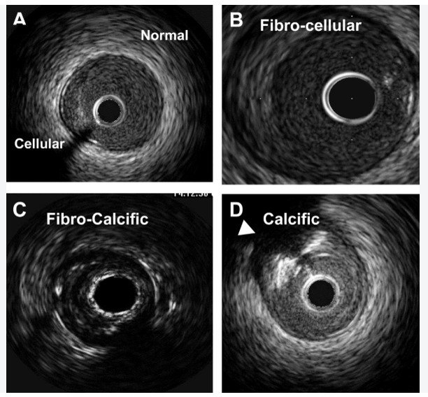
IVUS (intravascular ultrasound) means imaging the coronary artery wall from the inside, by advancing a thin catheter with an ultrasound transducer at its tip into the arteries that supply the heart. Coronary angiography gives a two-dimensional shadow image of a three-dimensional vessel. The clinical consensus statement of the European Association of Percutaneous Cardiovascular Interventions (EAPCI) and the European Bifurcation Club lists the limitations of angiography as "low spatial resolution and frequent branch overlaps", and states that in bifurcation lesions angiography underestimates the complexity. IVUS shows the inside of the vessel directly.
When is it used?
The 2024 ESC Guidelines for the management of chronic coronary syndromes recommend that PCI on anatomically complex lesions - in particular left main stem, true bifurcations and long lesions - be performed with intracoronary imaging guidance by IVUS or OCT, in the guideline's strongest recommendation category (class I, level of evidence A). The same guideline states that IVUS should be considered to assess the severity of intermediate left main stenosis (visually 40-70%) before revascularisation (class IIa, level of evidence B). The 2025 ACC/AHA acute coronary syndrome guideline likewise recommends intracoronary imaging in patients undergoing coronary stent implantation in the left main artery or in complex lesions (class 1, level of evidence A); this recommendation was upgraded from the lower class in the 2021 revascularisation guideline on the grounds of better stent expansion and less coronary dissection.
How is it done?
IVUS is not a separate procedure; it is performed over the guide catheter and guidewire already in use during the intervention. Unless contraindicated, intracoronary nitroglycerin is given before the catheter is advanced; when imaging is finished the catheter is removed and a final angiogram is taken to confirm that the vessel has not been injured. IVUS does not require the blood to be cleared from the vessel to obtain images; in this respect it differs from OCT, which needs contrast injection to displace blood. The EAPCI document notes that, because it allows working with a lower contrast volume, IVUS may be preferred in patients at high risk of contrast-associated acute kidney injury.
What does it show the physician?
- The true diameter and dimensions of the vessel
- The extent and composition of plaque (soft, fibrous or calcified plaque appears with different density)
- Vessel remodelling, the presence of thrombus and dissection, and the distinction between true and false lumen
This information is used to select the appropriate balloon and stent size and, at the end of the procedure, to see that the stent is adequately expanded and apposed to the vessel wall. The EAPCI document also defines criteria for a good result numerically (for example a minimum stent area above 5.5 mm² in non-left main lesions). These values are intended for the physician's decisions during the procedure.
Benefits and limitations
In the RENOVATE-COMPLEX-PCI trial reported in the 2024 ESC guideline (1639 patients), the composite of cardiac death, target vessel-related myocardial infarction or target vessel revascularisation within two years was 7.7% with intracoronary imaging guidance and 12.3% with angiography guidance alone (HR 0.64; 95% CI 0.45-0.89). This is an average and does not show what the outcome will be in an individual patient. IVUS does not measure how much a narrowing limits blood supply to the heart; pressure measurements such as FFR/iFR or stress imaging are used for that, and the sources state that IVUS should not be used on its own to justify an intervention. Imaging adds procedure time and cost, and some dissections may not be seen on IVUS. On safety, the EAPCI document describes imaging-related complications as "exceedingly rare"; in a registry covering more than 3600 procedures the rate was 0.6%, and the events were self-limiting or easily treated. The reported major complication rate is 0.1-0.5%, and the most frequently reported complication is coronary artery spasm (2.9%).
When should you contact your doctor?
Whether intracoronary imaging is needed, and the choice between IVUS and OCT, are decisions for the physician treating you. If you have had a stent implanted and your symptoms have returned, contact your own doctor: the 2024 ESC guideline encourages intracoronary imaging to identify the cause in stent thrombosis and in in-stent restenosis, and treatment differs according to the cause found. The information on this page is for general information and does not replace your own doctor's advice for your situation.
Sources
This page has been compiled from the guidelines and publications below. It is for information only and does not replace your physician's advice.
- European Society of Cardiology (ESC) -- 2024 ESC Guidelines for the management of chronic coronary syndromes, European Heart Journal 2024 (doi:10.1093/eurheartj/ehae177), Tablo: Assessment o
- European Society of Cardiology (ESC) -- 2024 ESC Guidelines for the management of chronic coronary syndromes, European Heart Journal 2024
- American College of Cardiology / American Heart Association -- 2025 ACC/AHA/ACEP/NAEMSP/SCAI Guideline for the Management of Patients With Acute Coronary Syndromes, Circulation / JACC 2025
- European Society of Cardiology (ESC) -- 2024 ESC Guidelines for the management of chronic coronary syndromes, bölüm 4.4.5.6 (RENOVATE-COMPLEX-PCI, Lee ve ark. aktarımı)
- Ahmed ve ark. -- Intravascular Ultrasound-Guided Versus Angiography-Guided Percutaneous Coronary Intervention: A Systematic Review, Meta-Analysis, and Meta-Regression of Randomized Control T
- EAPCI (ESC) ve European Bifurcation Club -- Intracoronary imaging for left main percutaneous coronary intervention: a clinical consensus statement, European Heart Journal, 2025
- American College of Cardiology -- Coronary Interventions Handbook, Chapter 12: Step-by-Step Technique for Intravascular Ultrasound (IVUS), Velagapudi P, Abbott JD
- American College of Cardiology -- Coronary Interventions Handbook, Chapter 12: Step-by-Step Technique for Intravascular Ultrasound (IVUS)
- EAPCI (ESC) -- Clinical use of intracoronary imaging. Part 1: guidance and optimization of coronary interventions, expert consensus document, EuroIntervention / European Heart Journal, 2018
- EAPCI (ESC) ve European Bifurcation Club -- Intracoronary imaging for left main PCI clinical consensus statement, European Heart Journal 2025; sol ana koroner dışı eşik için ACC Coronary Int
Last updated: October 13, 2026