Pulmoner Kapak Darlığı
Pulmoner kapak, kalpte sağ karıncıktan çıkan akciğer atardamarında (Pulmoner arter) bulunan bir kapaktır.
The pulmonary valve is the semilunar valve located at the origin of the pulmonary artery, the great vessel that arises from the right ventricle and carries deoxygenated blood to the lungs. Deoxygenated blood returning from the body reaches the right atrium, passes into the right ventricle, and the right ventricle pumps it through the pulmonary artery to the lungs in order to be oxygenated. When a stenosis develops at the level of the pulmonary valve, the afterload of the right ventricle increases, and the right ventricle has to work at a higher pressure and with greater force to eject blood across the narrowed valve. Over time, this results in excessive hypertrophy and strain of the right ventricular myocardium and, ultimately, in right heart failure.
Isolated pulmonary stenosis is almost always a congenital heart disease, and it accounts for 7-10% of all congenital heart diseases. Very rarely in adults, carcinoid plaques, cardiac tumours and a large sinus of Valsalva aneurysm may produce pulmonary stenosis through external compression.
In adults, pulmonary stenosis is classified into three main types according to the level of the obstruction:
- Stenosis at the level of the valve itself (valvular pulmonary stenosis)
- Stenosis below the valve (subvalvular, that is, infundibular pulmonary stenosis)
- Stenosis above the valve (supravalvular pulmonary stenosis)
In some cases, pulmonary stenosis may be accompanied by intracardiac shunts such as an atrial septal defect (ASD) or a ventricular septal defect (VSD), or by other congenital cardiac anomalies such as tetralogy of Fallot, transposition of the great arteries and double outlet right ventricle.
The most common form of the disease is valvular pulmonary stenosis, in which the obstruction involves the valve itself. In valvular pulmonary stenosis the leaflets are thickened and rigid, the cusps are deformed and fused to one another, giving the valve a domed appearance. For this reason the valve cannot open completely during right ventricular systole, and the right ventricle begins to work at a higher pressure and with greater force.
Beyond the neonatal period, valvular pulmonary stenosis frequently remains asymptomatic in children unless the obstruction is severe. In adulthood, as a consequence of the increase in right ventricular mass and of right ventricular myocardial dysfunction, dyspnea, dizziness and syncopal attacks begin to appear. In less severe stenosis, the only finding may be a murmur heard on cardiac auscultation. In adults, conditions associated with an increased volume load, particularly pregnancy, may render the disease considerably more symptomatic. In some patients with valvular pulmonary stenosis the valve may be dysplastic; this form is frequently encountered in a specific disorder known as Noonan syndrome.
In the less common subvalvular form, there is a tube-like narrowing beneath the valve, in the region called the infundibulum, caused by an increase in muscle mass. In the supravalvular form, the narrowing may be situated in the mid portion of the pulmonary artery, at the bifurcation or in the distal branches. The supravalvular type may be seen in certain specific syndromes (Alagille syndrome, Williams syndrome) and may also develop as a consequence of congenital rubella infection.
The severity of pulmonary stenosis is assessed by the pressure gradient between the right ventricle and the pulmonary artery. In mild to moderate pulmonary stenosis, the right ventricular pressure remains lower than the left ventricular pressure, whereas in severe pulmonary stenosis the right ventricular pressure rises markedly and may exceed the left ventricular pressure. If the pulmonary stenosis is accompanied by an intracardiac defect, mixing of deoxygenated blood with oxygenated blood may give rise to cyanosis.
In pulmonary stenosis, treatment is determined by the level and the severity of the obstruction and by the presence or absence of accompanying congenital anomalies. In valvular pulmonary stenosis, in which the stenosis involves the valve itself, the primary recommended treatment is opening of the pulmonary valve with a balloon, known as the closed (percutaneous) method, whereas in the other types open surgical repair may be performed according to the severity of the disease.
How Is Pulmonary Valve Stenosis Diagnosed?
Today the diagnosis of pulmonary valve stenosis can readily be established with the highly advanced ultrasound systems used for echocardiography. Transthoracic echocardiography (TTE), performed simply by placing an ultrasound probe on the anterior chest wall, allows the structure of the pulmonary valve to be examined and shows whether doming or dysplasia of the valve is present; the pressure gradient is then calculated by Doppler echocardiography and the severity of the stenosis is determined.
Once the diagnosis of pulmonary stenosis has been made, transesophageal echocardiography (TEE), performed with a probe swallowed through the mouth and positioned in the esophagus, readily demonstrates the severity of the stenosis, whether any accompanying regurgitation is present, and whether the valve is domed or dysplastic; if balloon treatment is planned, the diameter of the pulmonary annulus can also be measured precisely. In addition to two-dimensional TEE, three-dimensional echocardiography, which displays valve morphology particularly clearly, allows the presence and severity of the stenosis to be assessed with ease. Echocardiography is therefore a highly useful diagnostic tool for establishing the diagnosis of pulmonary stenosis and for planning its treatment. In our clinic, TTE together with both two-dimensional and three-dimensional TEE is performed in every patient with suspected pulmonary stenosis.
How Is the Closed, Non-Surgical Treatment Performed?
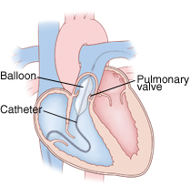
Balloon pulmonary valvuloplasty is the dilatation of the stenotic pulmonary valve with the aid of a balloon, without the need for surgery. In suitable patients it is the treatment of first choice today. The procedure is carried out in the angiography laboratory.
The veins of the groin carry deoxygenated blood from the legs to the right side of the heart through the inferior vena cava. The femoral vein is punctured with a small needle and a sheath is placed into the vein. Atraumatic wires and catheters that do not damage the vessel are advanced through this sheath into the right atrium, and the valve between the right atrium and the right ventricle is crossed to reach the right ventricle. With a dedicated guidewire or with the catheter itself, the stenotic pulmonary valve is crossed and the distal pulmonary artery is reached, and the pressure gradient between the right ventricle and the pulmonary artery is measured. A supportive stiff wire is then positioned in the distal pulmonary artery, and over this wire a dedicated balloon of appropriate diameter is advanced and seated across the stenotic pulmonary valve. The balloon is inflated briefly to separate the fused leaflets and to open the valve, so that the passage of blood to the lungs becomes easier and the pressure gradient across the valve decreases. After the procedure the pressure gradient is measured again and the valve is checked for adequate opening and for regurgitation; if the result is satisfactory, the procedure is terminated. The procedure generally takes 30-60 minutes, and most patients can be discharged the following day.
For Which Patients Is Balloon Treatment Recommended?
Balloon pulmonary valvuloplasty is performed particularly in children and adults who:
- have moderate or severe pulmonary valve stenosis;
- have complaints of dyspnea, early fatigue, chest pain or syncope;
- have a demonstrated increase in the load imposed on the right heart;
- have congenital pulmonary valve stenosis.
In some patients, even in the absence of marked complaints, treatment may be recommended if echocardiography demonstrates that the valve stenosis is severe.
The advantages of the treatment may be summarized as follows:
- it does not require open heart surgery;
- the success rate is very high;
- the hospital stay is short;
- recovery is rapid and the long-term results are highly favourable.
Can the Closed Method Be Applied to Every Patient?
The closed method can be applied to every patient with valvular pulmonary stenosis whose valve morphology is suitable for balloon dilatation. However, if the valve stenosis is extremely advanced, if other accompanying valvular diseases or congenital anomalies are present, or if the valve disease is accompanied by severe coronary artery disease, repair by open surgery may be undertaken instead.
Does the Balloon or the Procedure Carry Any Risks?
The complications of this closed treatment are far lower than those of open surgery. Complications that may arise during open surgery, such as serious bleeding, the consequences of prolonged anesthesia, and injury to vessels and cardiac chambers, are not frequently encountered. Moreover, because the procedure is performed without opening the chest, through no more than a small needle puncture and a sheath in the groin, recovery and the time to discharge are considerably shorter. Rupture of the balloon during the procedure, or injury caused by the stiff wire advanced into the distal pulmonary artery, may occur, but such complications are exceedingly rare in experienced hands compared with open surgery. For this reason, in suitable patients both the European Society of Cardiology and the American Heart Association recommend balloon pulmonary valvuloplasty as the first-line treatment for pulmonary valve stenosis.
The treatment decision is made by a cardiologist after clinical examination, echocardiography and any other investigations that may be required.
Last updated: October 13, 2026