Klasik Tanı Yöntemleri
Kalp damar hastalığı tanısı tek bir teste dayanmaz: şikâyetin özellikleri, yaş, cinsiyet ve risk faktörleri değerlendirilerek hastalık olasılığı tahmin edilir ve…
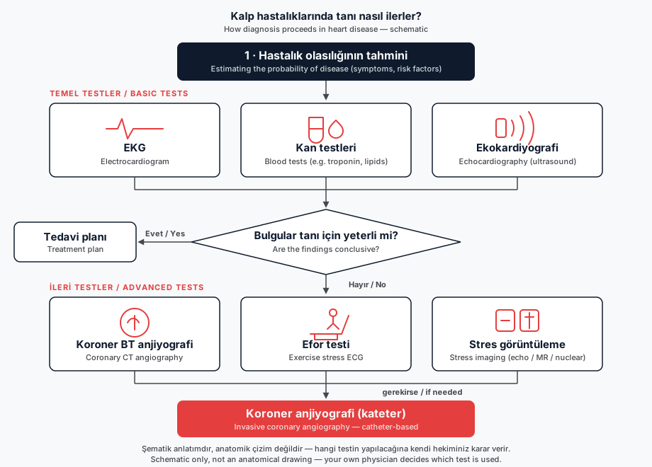
Diagnosis never rests on one test: symptom character, age, sex and risk factors are assessed first to estimate the likelihood of coronary disease, and tests are chosen accordingly (ESC Guidelines on chronic coronary syndromes, 2024). The 2021 AHA/ACC Chest Pain Guideline likewise selects tests after a structured risk assessment, with the patient part of the decision; low-risk patients need no urgent testing.
First step: ECG, blood tests, echocardiography
A resting 12-lead ECG is recommended in everyone reporting chest pain unless a clear non-cardiac cause is found; one recorded during or just after an episode is most valuable (ESC 2024; class I, level C). Blood tests recommended for all patients: lipid profile with LDL cholesterol, full blood count, creatinine, HbA1c and/or fasting glucose, thyroid function; high-sensitivity troponin if acute myocardial injury is suspected.
Resting echocardiography is recommended too (class I, B): it measures left ventricular contractile and diastolic function and detects wall motion abnormalities, hypertrophy, valve disease or pericardial effusion. If it is inconclusive, cardiac magnetic resonance may be considered (class IIb, C).
Which further test
ESC 2024 estimates likelihood from sex, age, angina character and risk factors: no testing at very low (≤5%) likelihood, uncertain benefit at low (>5–15%), and testing recommended from moderate (>15–50%) upwards. The first test depends on patient characteristics and local expertise and equipment (ACC, 2024).
- Coronary CT angiography shows the arteries themselves; recommended at low to moderate (>5–50%) likelihood (class I, A). Its dose is 3–5 mSv (CAR/CSTR, 2024); annual natural background radiation in the US is about 3 mSv (SCCT, 2022). A coronary calcium score of zero has a negative predictive value above 95% for obstructive disease, although in younger patients obstructive disease can be present without calcification (ESC 2024). It is not recommended in severe renal failure, decompensated heart failure, heavy calcification or an irregular rhythm (class III, C).
- Exercise ECG has 58% sensitivity and 62% specificity for obstructive disease, so it mainly assesses exercise tolerance, symptoms, arrhythmia, blood pressure response and event risk (class I, C).
- Stress echocardiography is recommended from moderate to high (>15–85%) likelihood (class I, B); it uses no radiation and can be repeated, but is operator dependent.
- Perfusion imaging with SPECT or PET shows reduced myocardial blood flow and scar (class I, B); stress is induced by exercise, or pharmacologically in those unable to exercise.
- Stress cardiac magnetic resonance is also recommended (class I, B): no radiation, but longer scanning, and not possible with a non-compatible pacemaker.
Preparation and angiography
Preparation differs between centres; the instructions you receive apply. Guideline points: no food for 3 hours before an exercise test, comfortable clothing and walking shoes (AHA, 2013); no solid food 3–4 hours before coronary CT, heart rate usually lowered to 60/min or below (SCCT, 2016); caffeine stopped at least 12 and preferably 24 hours before pharmacological perfusion imaging, with a 4-hour fast before the scan (water allowed) (SNMMI/EANM/ASNC/ACNM, 2025). Medication changes are decided only by your own physician.
Invasive coronary angiography is recommended at very high (>85%) likelihood, with severe symptoms refractory to medical therapy, or with high event risk (class I, C); radial (wrist) access is recommended (class I, A). It is not risk-free: major complications are reported in 0.5–2.0% after diagnostic femoral catheterisation, and death, infarction or stroke in about 0.1–0.2% with radial access (ESC 2024). Whether an intermediate narrowing really limits flow is measured with a pressure wire (FFR ≤0.80, iFR ≤0.89).
According to the guidelines the most valuable ECG is the one recorded during or just after an episode, so an assessment made while symptoms are still present gives the physician more information. Symptoms that persist despite treatment, or that appear at progressively lower levels of exertion, are situations the guidelines consider important for further assessment; this information belongs with your own physician, who decides which test is appropriate.
Sources
This page has been compiled from the guidelines and publications below. It is for information only and does not replace your physician's advice.
- European Society of Cardiology (ESC) – 2024 ESC Guidelines for the management of chronic coronary syndromes, Öneri Tablosu 1 / Bölüm 3.1.2.1 (2024)
- European Society of Cardiology (ESC) – 2024 ESC Guidelines for the management of chronic coronary syndromes, Öneri Tablosu 4 (2024)
- European Society of Cardiology (ESC) – 2024 ESC Guidelines for the management of chronic coronary syndromes, Bölüm 3.2.3 (2024)
- European Society of Cardiology (ESC) – 2024 ESC Guidelines for the management of chronic coronary syndromes, Öneri Tablosu 5 (2024)
- American Heart Association (AHA) – Exercise Standards for Testing and Training: A Scientific Statement from the American Heart Association, Circulation (2013)
- European Society of Cardiology (ESC) – 2024 ESC Guidelines for the management of chronic coronary syndromes, Bölüm 3.2.1 ve Öneri Tablosu 3 (2024)
- European Society of Cardiology (ESC) – 2024 ESC Guidelines for the management of chronic coronary syndromes, Öneri Tablosu 8 (2024)
- European Society of Cardiology (ESC) – 2024 ESC Guidelines for the management of chronic coronary syndromes, Öneri Tablosu 8 ve Bölüm 3.3.1.3 (2024)
- Society of Cardiovascular Computed Tomography (SCCT) – SCCT guidelines for the performance and acquisition of coronary computed tomographic angiography, Journal of Cardiovascular Computed To
- Society of Cardiovascular Computed Tomography (SCCT) – 2022 Use of coronary computed tomographic angiography for patients presenting with acute chest pain to the emergency department: SCCT e
Last updated: October 13, 2026