Shockwave Lithotripsy
Şiddetli kalsifik koroner lezyonların şok dalgası teknolojisiyle yumuşatılarak stent yerleşimine hazırlanması.
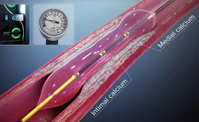
Shockwave intravascular lithotripsy (IVL) is a method that breaks up the calcium deposited in the wall of the heart arteries using acoustic pulses. Emitters mounted inside the balloon at the tip of the catheter generate acoustic pressure waves within the fluid filling the balloon; these waves fracture the calcium in the vessel wall, after which the narrowing can be dilated with low balloon pressure. No tissue is cut, shaved or removed with this method (Shockwave Medical C2 coronary IVL catheter instructions for use, November 2025 revision).
The device's approved indication is lithotripsy-enabled, low-pressure balloon dilatation of severely calcified, stenotic, de novo coronary arteries before stent placement. The device is not designed to deliver a stent, and its use in the carotid (neck) or cerebral vessels is contraindicated, meaning it is not suitable there (same instructions for use, November 2025 revision).
Why calcification matters
Roughly 25–30% of patients undergoing angiography or stenting have angiographically visible calcification in the vessel wall; the rate detected by intravascular imaging (IVUS/OCT) is almost twice the angiographic rate. Calcification is most often associated with older age, male sex, hypertension, diabetes and renal failure (Interventional Cardiology Review 2025 review article).
The core problem is that insufficiently modified plaque prevents full stent expansion and leaves a small final stent area. In patients with angiographic calcification, 1-year target lesion failure has been reported as 8.7% versus 6.0% and major adverse cardiac events as 19.9% versus 12.9% (pooled analysis of the HORIZONS-AMI/ACUITY trials, as reported in the Interventional Cardiology Review 2025 review). The same review cites stent expansion above 80% and a minimum stent area of 5.5 mm² by IVUS or 4.5 mm² by OCT as targets.
When it is considered
The decision rests on imaging. The 2023 clinical consensus statement of EAPCI, the interventional branch of the European Society of Cardiology (European Heart Journal 2023), recommends intravascular imaging and/or advanced plaque modification rather than more aggressive balloon inflation when a stenosis cannot be dilated with a semi-compliant or high-pressure balloon. The same statement lists the settings in which IVL is appropriate:
- deep calcification and calcified nodules,
- large-diameter vessels,
- stent underexpansion (this indication is currently off-label),
- bifurcation lesions in which the side branch must be kept wired,
- aorto-ostial calcified stenoses.
The 2024 ESC guidelines for the management of chronic coronary syndromes likewise recommend that the procedure be performed under IVUS or OCT guidance in anatomically complex lesions (left anterior descending artery, true bifurcations, long lesions).
How the procedure is performed
The values below are those stated in the device's November 2025 instructions for use. Balloon diameter is sized 1:1 to the vessel diameter; when an exact 1:1 size is unavailable, the largest available balloon is used. The balloon must be advanced across and positioned at the target stenosis, so lesions the balloon cannot cross represent a limit of the method. During lithotripsy the balloon is inflated to no more than 4 atmospheres; the pulse rate is 1 pulse per second; a maximum of 10 consecutive pulses is delivered, after which the device enforces a pause of at least 10 seconds. The limit of 80 pulses per treatment site is not exceeded. Catheters are available in 2.5–4.0 mm diameters with a 12 mm balloon length.
Results and possible risks
In the pivotal DISRUPT CAD III study (384 patients; data published in the Journal of the American College of Cardiology 2020 and reported in the device's November 2025 instructions for use), freedom from major adverse cardiac events (cardiac death, myocardial infarction or target vessel revascularisation) at 30 days was 92.2% and procedural success was 92.4%; a stent could be delivered in 99.2% of patients. The overall 30-day event rate was 7.8%: cardiac death 0.5%; non-Q-wave myocardial infarction (mostly the in-hospital type detected by blood tests) 6.0%; Q-wave myocardial infarction 1.6%; target vessel revascularisation 1.6%; coronary perforation 0.3%. Severe dissection (type D–F) occurred in no patient.
One point specific to this method is that the mechanical pulses generated may cause extra beats in patients with a slow heart rate and may affect the sensing function of a permanent pacemaker or defibrillator. For this reason heart rhythm and blood pressure are monitored continuously throughout the procedure, and treatment is paused temporarily if a notable effect is seen. In DISRUPT CAD III no serious adverse event related to this warning was observed.
How it differs from rotablation
In rotational atherectomy a diamond-coated burr rotates at high speed, preferentially abrading rigid, non-elastic tissue and removing calcium from the vessel, whereas IVL fractures calcium in place. The 2023 EAPCI consensus statement notes that IVL may be preferred for deep calcium while atherectomy may be more effective for superficial calcium, but it also states explicitly that data directly comparing the efficacy of the two are still lacking. In the small, unblinded ROTA.shock trial (70 patients; Catheterization and Cardiovascular Interventions 2023), mean minimum stent area was 6.10 mm² with IVL and 6.60 mm² with rotational atherectomy; because the difference did not exceed the pre-specified margin, IVL was considered non-inferior to rotational atherectomy.
Your own physician makes the decision
Whether this method is suitable for your own vessel anatomy is assessed by the physician following you, based on angiographic and intravascular imaging findings. Your pre- and post-procedural follow-up plan, the medications you need to take, and what to do if symptoms develop vary from person to person; please direct these questions to your own physician.
Sources
This page has been compiled from the guidelines and publications below. It is for information only and does not replace your physician's advice.
- Shockwave Medical – Shockwave C2 Coronary IVL Catheter, Instructions for Use (Kullanım Talimatı), LBL 63927 Rev G, Kasım 2025 – 'Indications for Use' ve 'Contraindications for Use' bölümleri
- Shockwave Medical – Shockwave C2 Coronary IVL Catheter IFU, LBL 63927 Rev G, Kasım 2025 – 'Device Description' bölümü
- Shockwave Medical – Shockwave C2 Coronary IVL Catheter IFU, LBL 63927 Rev G, Kasım 2025 – 'Device Description', 'Treating the Site with Intravascular Lithotripsy' ve teknik özellikler tablos
- Shockwave Medical – Shockwave C2 Coronary IVL Catheter IFU, LBL 63927 Rev G, Kasım 2025 – 'Precautions' madde 4 ve işlem basamakları
- Barbato E, Gallinoro E, Abdel-Wahab M, ve ark. 'Management strategies for heavily calcified coronary stenoses: an EAPCI clinical consensus statement in collaboration with the EURO4C-PCR grou
- Barbato E, ve ark. EAPCI clinical consensus statement, European Heart Journal 2023;44(41):4340–4356 – rotasyonel aterektomi bölümü ve uzlaşı ifadesi 12
- Barbato E, ve ark. EAPCI clinical consensus statement, European Heart Journal 2023;44(41):4340–4356
- Blachutzik F, ve ark. 'Coronary intravascular lithotripsy and rotational atherectomy for severely calcified stenosis: Results from the ROTA.shock trial.' Catheterization and Cardiovascular I
- Shockwave C2 Coronary IVL Catheter IFU, LBL 63927 Rev G, Kasım 2025 – 'Clinical Studies', Tablo 4 ve Tablo 6 (DISRUPT CAD III); aynı veriler: Hill JM, ve ark. Journal of the American College
- Shockwave C2 Coronary IVL Catheter IFU, LBL 63927 Rev G, Kasım 2025 – Tablo 5, anjiyografik komplikasyonlar tablosu ve 'Warnings' madde 11
Last updated: October 13, 2026