Sol Atriyal Apendiks Trombozu
Sol atriyal apendiks (LAA), kalbin sol kulakçığına bağlı küçük bir kesedir. Bu kesenin içinde kan pıhtısı (trombüs) oluşmasına sol atriyal apendiks trombozu denir.
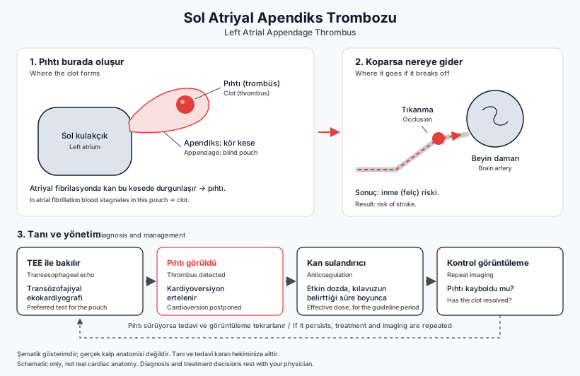
The left atrial appendage (LAA) is a small pouch attached to the left atrium of the heart. When a blood clot (thrombus) forms inside this pouch, it is called left atrial appendage thrombus. If the clot breaks loose, it can travel through the circulation to the brain and cause a stroke, which is why this finding is taken seriously.
Why does it form, and in whom?
The most common underlying condition is the rhythm disorder called atrial fibrillation (AF). The 2024 ESC Guidelines for the management of atrial fibrillation state that, left untreated, the risk of ischaemic stroke in AF is increased five-fold and that one in every five strokes is associated with AF. In non-rheumatic AF, the great majority of clots forming in the left atrium (reported as approximately 90% in peer-reviewed reviews) are located inside this pouch.
The reason lies in the structure of the appendage: its distinctive shape and the trabeculations (ridge-like muscle bundles) on its inner surface cause blood stasis within the pouch, and its junction with the atrium is narrow. In atrial fibrillation, flow within the appendage turns into a rapid, shallow filling-and-emptying cycle with lower flow velocities. This low-velocity pattern is the flow pattern most strongly associated with "spontaneous echo contrast" on echocardiography and with clot formation. In pooled analyses, the factors that increase the likelihood of thrombus are non-paroxysmal (persistent or permanent) AF (OR 3.6; 95% CI 2.4-5.2), previous stroke (OR 3.0), hypertension (OR 2.9) and heart failure.
How is it diagnosed?
Standard (transthoracic) echocardiography does not reliably visualise this pouch, so it is not sufficient to rule out a clot. The preferred method is transoesophageal echocardiography (TOE/TEE), in which the heart is imaged from behind using an ultrasound probe passed down the oesophagus; reviews report an accuracy of 99.1% and a negative predictive value of 98.9% for detecting atrial thrombus. Cardiac computed tomography with a delayed-contrast protocol has emerged as an alternative method: its sensitivity and negative predictive value are high while its positive predictive value is low, so it is reliable for ruling a clot out, whereas a positive result requires confirmation with TOE.
Taking an anticoagulant is not by itself proof that no clot is present. In a pooled analysis of 56,660 patients with AF, atrial thrombus was detected on pre-cardioversion TOE in 7.5% of patients not on anticoagulation, 5.5% of those on anticoagulation and 4.9% of those on adequately dosed anticoagulation; before catheter ablation the corresponding rates were 1.8%, 1.8% and 1.3%.
What happens if a clot is found?
According to the 2024 ESC guideline, at least 3 weeks of effective anticoagulation should be given before cardioversion (adherence to a DOAC, or an INR above 2 if a vitamin K antagonist is used); if this has not been achieved, TOE is recommended to exclude intracardiac thrombus (Class I). When AF has lasted longer than 24 hours, early cardioversion is not recommended unless one of these two conditions is met.
- If a clot is seen, cardioversion is deferred: the 2024 ESC guideline states that therapeutic anticoagulation should be instituted for a minimum of 4 weeks, followed by repeat TOE to confirm that the thrombus has resolved; the 2023 ACC/AHA/ACCP/HRS guideline recommends at least 3 to 6 weeks of therapeutic anticoagulation, after which imaging should be repeated (Class 1, C-LD).
- The clot resolves in most patients, but not in every patient, and it can take weeks: resolution on follow-up TOE was reported in 61.2% after 3-12 weeks of a vitamin K antagonist (CLOT-AF registry) and in 41.5% after 6 weeks of rivaroxaban (X-TRA; 60.4% when resolution or reduction in size were counted together).
- If a clot is found while the patient is already taking an anticoagulant, adherence, dose adequacy, drug absorption and drug interactions are assessed. Switching to an alternate anticoagulant can be considered, although, in the guideline's own words, the benefit is uncertain.
Device closure of the left atrial appendage is not performed while an intracardiac clot is present; the manufacturer's official instructions for use list intracardiac thrombus as a contraindication, because moving the catheter inside the pouch could dislodge the clot. The procedure comes into consideration only after the clot has resolved, and in the guidelines it is an option that may be considered for patients who cannot take long-term anticoagulation (2024 ESC: Class IIb, level of evidence C).
When should you contact your doctor?
Do not stop your anticoagulant or change its dose on your own; if you are having difficulty taking it, tell your doctor, because dose and adherence affect whether the clot resolves. Be sure to attend the follow-up imaging that has been planned for you. If a sudden symptom suggesting a stroke develops, go to an emergency department without delay. The information on this page is general and does not replace individual medical advice; which imaging and which treatment are appropriate for your situation is decided by the doctor who examines you.
Sources
This page has been compiled from the guidelines and publications below. It is for information only and does not replace your physician's advice.
- European Society of Cardiology (ESC) / EACTS — 2024 ESC Guidelines for the management of atrial fibrillation, Bölüm 6 ([A] Avoid stroke and thromboembolism), 2024
- Hakemli derleme — "The Left Atrial Appendage and Atrial Fibrillation — A Contemporary Review", J Clin Med / PMC10650862, 2023 (kendi kaynağı olarak Regazzoli D. ve ark., Biomed Res Int 2015'
- Hakemli derleme — "The Left Atrial Appendage and Atrial Fibrillation — A Contemporary Review", J Clin Med / PMC10650862, Bölüm 1 ve 2.2, 2023
- Hakemli derleme — "The Left Atrial Appendage and Atrial Fibrillation — A Contemporary Review", J Clin Med / PMC10650862, Bölüm 4, 2023
- Hakemli meta-analiz — Noubiap JJ ve ark., "Atrial thrombus detection on transoesophageal echocardiography in patients with atrial fibrillation undergoing cardioversion or catheter ablation:
- Hakemli meta-analiz — Noubiap JJ ve ark., J Cardiovasc Electrophysiol 2021;32:2179-88
- Hakemli derlemeler — "The Left Atrial Appendage and Atrial Fibrillation — A Contemporary Review" (PMC10650862, Bölüm 5/5.1), 2023 ve "Overcoming barriers for left atrial appendage thrombus:
- ESC/EACTS — 2024 ESC Guidelines for the management of atrial fibrillation, Bölüm 7.2.1 ve Öneri Tablosu 16 (Elektriksel kardiyoversiyon), 2024
- ESC/EACTS — 2024 ESC Guidelines for the management of atrial fibrillation, Bölüm 7.2.1, 2024
- ACC/AHA/ACCP/HRS — 2023 Guideline for the Diagnosis and Management of Atrial Fibrillation, Bölüm 8.2.1 (Prevention of Thromboembolism in the Setting of Cardioversion), Öneri 2 ve 3, Circulat
Last updated: October 13, 2026