CARDIOVASCULAR DISEASES
AORT COARCTATION
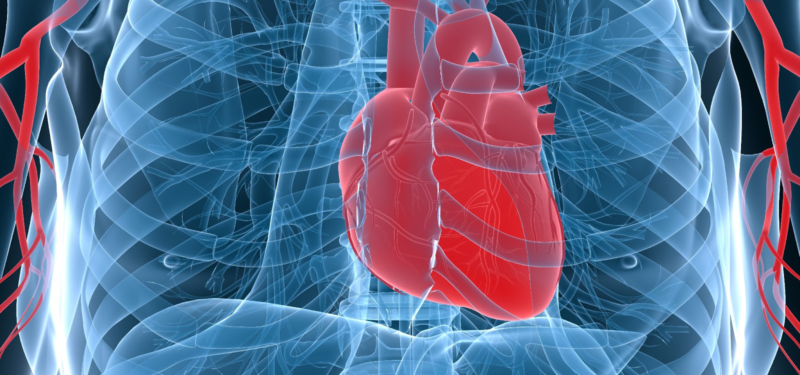
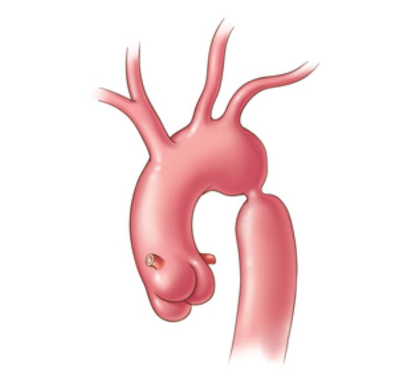
Aorta is the main vessel which carries oxygenated blood from left ventricle to the whole body. Aortic coarctation is a congenital condition whereby the aorta narrows in the area where the ductus arteriosus (ligamentum arteriosum after regression) inserts. Just like the aortic valve stenosis, afterload of the left ventricle incerases and left ventricular muscle mass increases in patients with aortic coarctation. If it is not diagnosed, left ventricular failure can develop. However, the most important symptomatic situation in aortic coarctation is hypertension in upper extremity arteries. On the other hand, blood pressure is decreased in lower extremity arteries because of the narrowing at the level of left subclavian branch of the aorta. Due to the different oxygenation of lower and upper extremities, power of upper and lower extremities are different and growth retardation can develop in lower extremities. The narrowing of the aorta can be asymptomatic and the diagnose can be incidentally during measurement of blood pressure or hearing a murmur on auscultation. In a normal person, blood pressure at legs is usually higher than arms. However, in aortic coartation, blood pressure in legs is decreased. Headache or epistaxis can bee seen in the patient due to hypertension.
HOW TO DIAGNOSE?
During physical examination, a murmur is usually heard and the difference between blood pressure in arms and legs is present. Echocardiography is an important diagnostic tool for the diagnosis of aortic coarctation. Transthorasic echocardiography (TTE) can demonstrate the coarctation. However, aortography is the ideal method both for the diagnosis and to determine the level of coarctation. It is also possible to measure the pressure gradients in cath lab. CT or MRI is also very useful diagnostic tools. In our clinic, we used TTE as a first diagnostic tool and then, the definite diagnosis is made by CT. Then, we perform catheterization and aortography for determining the therapy.
How do we apply percutaneous transcatheter treatment of aortic coarctation?